Branchiostoma lanceolatum
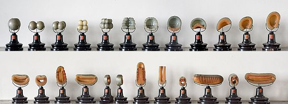
- Ziegler Wax model of Branchiostoma lanceolatum (around 1882)
Ziegler Wax Models
The model series by Adolf Ziegler (1820 -1889) of different development stages of the lancet fish Branchiostoma (Amphioxus) lanceolatum (Pallas, 1774) (redescribed by Poss & Boschung [1]) were substantially supported by the work of Berthold Hatschek (1854-1941). Ziegler’s studio for scientific sculpture, founded in Freiburg i.Br. in 1852, supplied university institutes with zoological, embryological and human anatomical models. The figures show the furrow divisions, blastula, gastrula and the development of the cotyledons.
Still under debate, the lancet fish is regarded, due to numerous similarities with vertebrates, foremost the physique of this animal and its embryonic development, as the closest living relative of vertebrates, which is why it attracted the interest of researchers very early on. By studying his embryonic development, Hatschek gained important insights into the formation and evolution of vertebrates [2,3]. Therefore, the model organism Amphioxus is the inspiration for comparative embryology as the basis of evolutionary biology of the 19th century (compare [4]) until now (see. [5,6])
The series shown is therefore still an important demonstration object today and has been used for more than 100 years in the training of young biologists. The material is a coloured beeswax mixture [7], first molded freehand and consecutively novated by the use of plaster molds. These techniques found even greater appeal with the use of microtomes and wax plates (see [8]).
References
- Poss, S. G., & Boschung, H. T. (1996). LANCELETS (CEPHALOCHORDATA: BRANCHIOSTOMATTDAE): HOW MANY SPECIES ARE VALID?. Israel Journal of Zoology, 42(sup1), S13-S66.
- Hatschek, B. (1884). Mittheilungen über Amphioxus. Zool. Anz, 7(177), 517-520.
- Hatschek, B. (1892). Die Metamerie des Amphioxus und des Ammocoetes. Anatomischer Anzeiger, 7, 454-464.
- Sander, K. (2012). Landmarks in developmental biology 1883–1924: historical essays from Roux’s Archives. Springer Science & Business Media.
- Onai, T. (2018). The evolutionary origin of chordate segmentation: revisiting the enterocoel theory. Theory in Biosciences, 1-16.
- Holland, N. D. (2017). The long and winding path to understanding kidney structure in amphioxus—a review. Int J Dev Biol, 61, 683-8.
- Hopwood, N. (2003). Embryos in wax: models from the Ziegler studio.
- Doll, S., Kirsch, J., & Eckart, W. U. (2017). Wenn der Tod dem Leben dient-Der Mensch als Lehrmittel: Institut für Anatomie und Zellbiologie. Springer-Verlag.


